HKU researchers resolve century-long debate about how bone cells develop
06 Aug 2014
The ability to walk and run is usually taken for granted but skeletal diseases such as osteoarthritis and osteoporosis have significant economic and human impact. As people live longer, these disorders are forecast to reach unprecedented levels globally and understanding how bones form and repair is very important for the development of therapies for bone diseases. Recently, researchers from Department of Biochemistry, Li Ka Shing Faculty of Medicine, The University of Hong Kong (HKU) have discovered that during bone formation and growth, cartilage cells (chondrocytes) survive and become bone cells (osteoblasts), overturning current dogma, resolving a century-long controversy. The discovery, published in the international prestigious journal, Proceedings of the National Academy of Sciences of USA, is important for understanding bone diseases.
Research Implications
Professor Kathryn Cheah, Jimmy and Emily Tang Professor in Molecular Genetics and Chair Professor of Department of Biochemistry, Li Ka Shing Faculty of Medicine, HKU, who led the study, points out that the discovery has far reaching implications for understanding bone growth and associated diseases and their treatment. “The discovery will spur researchers to revisit current concepts on the mechanism of skeletal disorders and has great importance for the development of treatment strategies for bone diseases such as osteoporosis.” Another researcher of the study, Professor Danny Chan, Professor of Department of Biochemistry, Li Ka Shing Faculty of Medicine, HKU adds, “Our study provides a new insight on cartilage and bone biology and we believe that descriptions of the process of bone formation in medical textbooks will have to be rewritten.”
Background of the study
In embryonic development most of the bones in the body develop in a multistep process in which stem cells condense and differentiate to form cartilage which acts as a template upon which bone is laid down. According to dogma, the chondrocytes and osteoblasts are two separate cell types that develop independently and it is generally accepted that death is the ultimate fate of chondrocytes. But whether a chondrocyte can change and become an osteoblast and contribute to the full range of the population of bone cells, has been the subject of a century-long debate.
Research method and findings
In the current study, the team used genetic methods to mark cartilage cells in mice with fluorescent protein and tracked their fate during bone development. They found that the labeled chondrocytes became osteoblasts in bones. This process occurs not only during embryonic development, but also in adulthood when bones are repairing after fractures.
About the research team
The research team was led by Professor Kathryn Cheah, Jimmy and Emily Tang Professor in Molecular Genetics and Chair Professor of Department of Biochemistry, Li Ka Shing Faculty of Medicine, HKU. Other researchers include, Professor Danny Chan, Dr Liu Yang, Dr Tsang Kwok-yeung and Ms Tiffany Tang of the Department of Biochemistry, Li Ka Shing Faculty of Medicine, HKU. The study was funded by grants from the University Grants Committee of Hong Kong and Hong Kong Research Grants Council.
To use the press release photo(s) for any publishing, publicity and related purpose, photo courtesy should be given to “Li Ka Shing Faculty of Medicine, The University of Hong Kong”

Group photo of the research team from Department of Biochemistry, Li Ka Shing Faculty of Medicine, The University of Hong Kong
(From left) Miss Tiffany Tang, MPhil student of Department of Biochemistry; Professor Kathryn Cheah, Jimmy and Emily Tang Professor in Molecular Genetics and Chair Professor of Department of Biochemistry; Professor Danny Chan, Professor of Department of Biochemistry and Dr Tsang Kwok-yeung, Post-doctoral Fellow of Department of Biochemistry
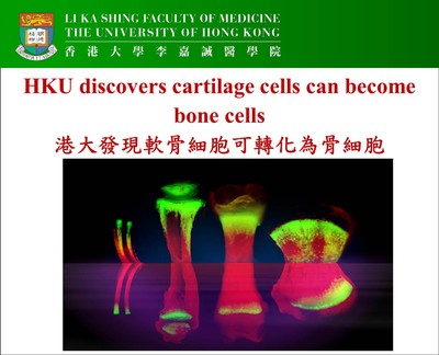
The picture shows different bones expressing fluorescent proteins. In the images in the lower part, cartilage cells (chondrocytes) have been genetically tagged to express green fluorescent protein. This represents the conventional view that cartilage cells (chondrocytes) and bone cells (osteoblasts) have different origins, with the former fated to death. As for the upper part, the images show bones in which cartilage cells (chondrocytes) were genetically labeled with fluorescent protein and their fate tracked during development. Unlike the lower images, bone cells are also labelled with fluorescent protein. This finding is the basis for the discovery that cartilage cells (chondrocytes) survive and become bone cells (osteoblasts) in bone formation.

The latest findings reveal that bone cells (osteoblasts), other than formed directly from bone stem cells, can also be originated from cartilage cells (chondrocytes) during bone formation and growth.