
Research Area 1: Diverse Imaging Research Activities at Imaging-Initiated Intervention and Intelligence Lab
- Discovery and development of novel imaging biomarkers for clinical applications in the design of treatment strategies for various diseases
- Assessment of high-precision quantitative and functional imaging methods to enable the design of clinical trials with reduced sample sizes or reduced follow-up
- Development of requisite imaging acquisition protocols and image analysis tools appropriate to the design of new treatment strategies
- Investigation of transformative, novel approaches guided by imaging that may lead to imaging-initiated minimally-invasive treatment methods
- Application of AI algorithms and machine learning techniques (deep learning, U-net segmentation, contrastive learning, recurrent convolution neural network, YOLO, natural language processing) to advance image-initiated clinical decision support programs that would improve the quality and efficiency of clinical work to address workforce shortages and increase job satisfaction
| Supervisor(s) |
|---|
|
Professor KT Bae |
Research Area 2: Molecular and Functional Imaging in Gynae-Oncology, Bridging the Imaging-Pathological Translation
- Body diffusion-weighted MR imaging (DW-MRI) − application in oncology, quantification of diffusion parameters and technique optimisation on 3T MRI
- DW-MRI quantification beyond mono-exponential model
- Qualitative and quantitative peritoneal imaging
- Application of radiomics in gynaecological oncology
| Supervisor(s) |
|---|
Research Area 3: Cardiac Magnetic Resonance (CMR) and Cardiac Computed Tomography (CCT)
- Stress cardiac magnetic resonance and perfusion mapping/quantification in
- Asymptomatic high risk diabetics coronary microvascular disease and empagliflozin
- CMR feature tracking for prognosis and diagnosis of diastolic dysfunction in heart failure with preserved ejection fraction and comparing this to catheter pressure measurements, echocardiography and CMR tagging
- 4D flow in heart failure with preserved ejection fraction
- Longitudinal study on CMR appearances of recovered COVID-19 patients, recovered viral respiratory infection patients, and post-vaccination patients
- Coronary artery plaque changes imaged on CCT in patients on empagliflozin
- Coronary artery volume and myocardial mass ratio for assessing coronary microvascular function imaged on CCT
- CT Coronary calcium scoring in different ethnicities
| Supervisor(s) |
|---|
Research Area 4: Advanced MRI and Deep Learning
- Deep-learning-based MRI reconstruction and quantification
- Deep-learning-based medical imaging segmentation and classification
- fMRI acquisition and modelling for brain function, BOLD and non-BOLD contrast mechanisms
- MRI pulse sequence design and novel imaging contrasts
- Molecular imaging, such as MR spectroscopy and chemical exchange saturation transfer
- MRI hardware development
| Supervisor(s) |
|---|
Research Area 5: Early Clinical Translatability of Advanced Imaging Techniques Leveraging Emerging Technologies and Complex Computational Methods
- Using imaging for early detection, cancer characteristics, and response assessment
- Nasopharyngeal carcinoma
- Hepatocellular carcinoma
- Prostate cancer
- Quantitative radiomics and radiogenomics
- Big data, machine learning, medical artificial intelligence
- Deep learning imaging applications to ageing and longevity
| Supervisor(s) |
|---|
|
Professor KT Bae |
Research Area 6: Advanced MRI and Other Molecular Imaging Techniques in Neurological Diseases
- Alzheimer's and other neurodegenerative diseases − volumetric MRI, functional MRI, MR perfusion, MR spectroscopy (MRS), amyloid and tau PET, susceptibility-weighted imaging (SWI), chemical exchange saturation transfer (CEST), artificial intelligence in dementia diagnosis
- Demyelinating diseases, such as systemic lupus erythematosus, neuromyelitis optica and multiple sclerosis − arterial spin labelling (ASL), diffusion tensor imaging (DTI), MRS
- Brain tumours − surrogate markers in anticancer drug trials using dynamic contrast enhanced (DCE) MR perfusion and MRS
- Epilepsy − volumetric MRI, T1 Rho, MRI/PET
- Stroke − MR perfusion (ASL) imaging in evaluation of stroke, MR vessel wall imaging in aneurysm, vasculitis and intracranial atherosclerosis, blood-brain-barrier imaging in haemorrhagic stroke, stroke imaging database in Hong Kong West Cluster of the Hospital Authority
| Supervisor(s) |
|---|
|
Professor KT Bae |
Find Out More
Information on selected projects or research areas: Interested candidates are advised to email the relevant supervisors. Please enclose with your email: (i) your CV, (ii) a brief description of your research interest and experience, and (iii) two reference letters (reference letters not required for HKUMed UG students seeking MRes[Med] or URIS projects)
Research studies enquiries specific to the Department/School’s research should be directed to the Departmental Research Postgraduate Advisor: Dr P Cao ( caopeng1@hku.hk)
Information on the research programme, funding support and admission requirements could be found on the RPg Admissions website.
General admission enquiries should be directed to rpgmed@hku.hk.
Meet Our Students
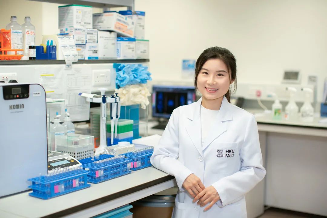
SHI Jingjing
Place of Origin: China
Progress: PhD Year 3
Supervisor: Prof PL Khong
史菁菁的首個運用小動物PET/MRI成像系統、以影像學為基礎的腫瘤研究。從對新領域一無所知,到摸索出有效的應用方法,從束手無策到得心應手。
目前動物的藥物應用研究廣泛,但真正轉化到臨床醫學的藥物極少,這其實是對基礎研究資源的巨大浪費。歸根結底,是由於對動物模型缺乏透徹了解。史菁菁表示,「我們的研究為幫助大家深刻認知動物模型的內在變化,以此提高臨床藥物應用的轉化率。」
史菁菁將堅定研究影像學領域,在臨床和科研同向發展,既成為一名影像科的醫生,同時從事動物成像及轉化醫學研究。「目前該領域還有很大的研究空間,希望盡我所能助推一步,而當醫生是在宏觀上助人的體現,幫助患者去解決病痛,這也是我的快樂來源。」
April 2021
Follow HKUMed