
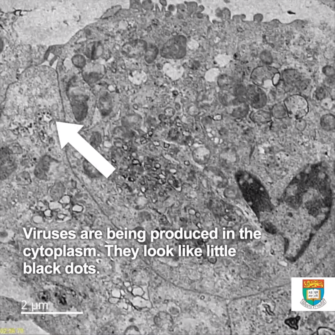
Thin-section electron micrographs of the 2019 novel coronavirus grown in cells at The University of Hong Kong. The image shows part of a virus infected cell grown in culture with multiple virus particles being released from the cell surface. Each infected cell produces thousands of new infectious virus particles which can go on to infect new cells.
Image credit: John Nicholls, Leo Poon and Malik Peiris, LKS Faculty of Medicine, and Electron Microscopy Unit, The University of Hong Kong

Pseudo-colour thin-section electron micrographs of the 2019 novel coronavirus grown in cells at The University of Hong Kong. The image shows part of a virus infected cell grown in culture with multiple virus particles being released from the cell surface. Each infected cell produces thousands of new infectious virus particles which can go on to infect new cells.
Image credit: John Nicholls, Leo Poon and Malik Peiris, LKS Faculty of Medicine, and Electron Microscopy Unit, The University of Hong Kong
Video of the new novel coronavirus growing in culture.
Video credit: John Nicholls, Leo Poon and Malik Peiris, LKS Faculty of Medicine, and Electron Microscopy Unit, The University of Hong Kong
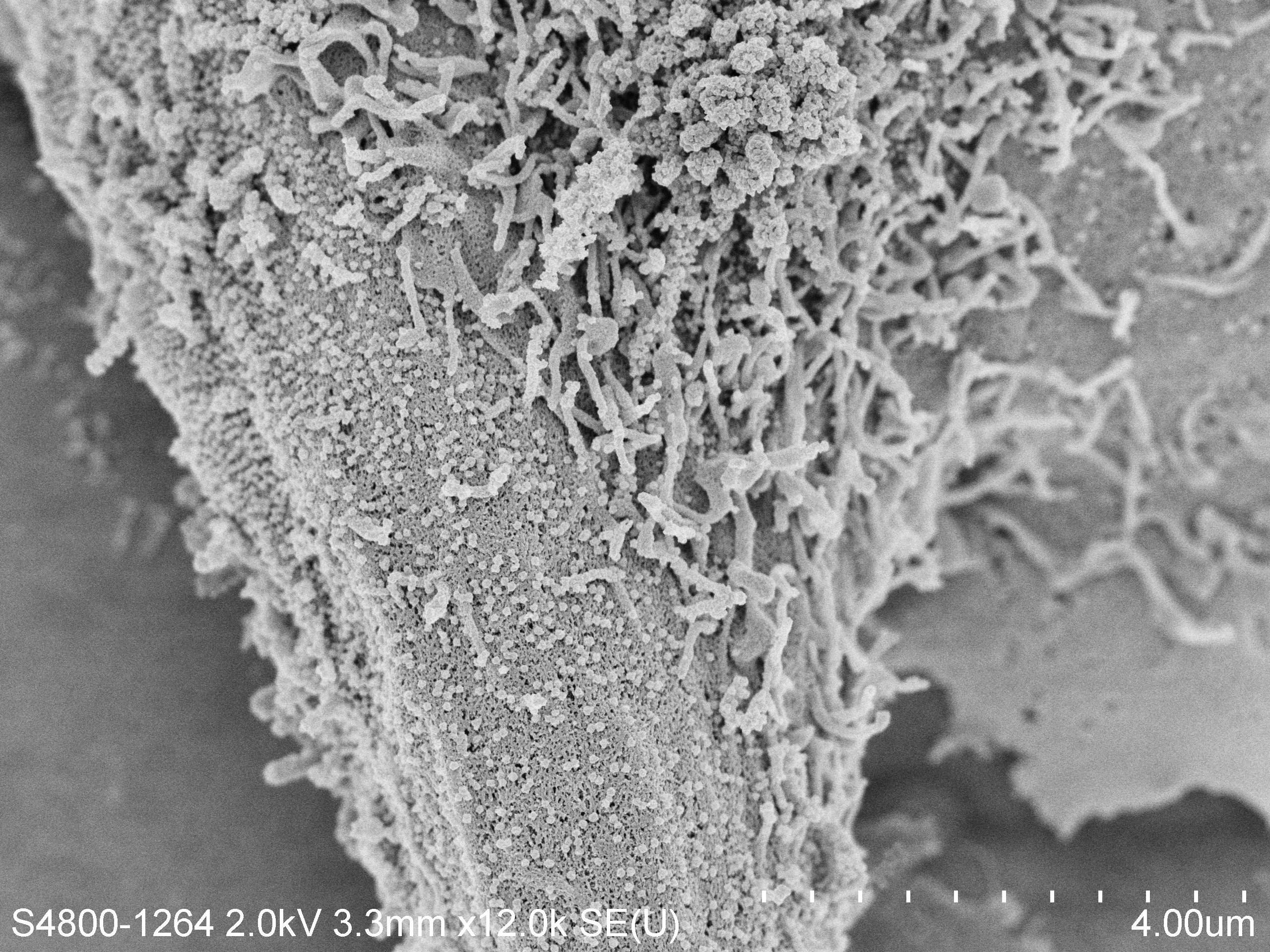
Scanning electron micrograph of coronavirus particles budding out from the surface of a cell culture after 24 hours of infection. The numerous small round spheres are the viral particle on the surface of the cell. The image is taken at a magnification of 24,000 times.
Image credit: LKS Faculty of Medicine, Department of Electrical and Electronic Engineering (K.Tsia, K.Lee and Q.Lai), and Electron Microscopy Unit, The University of Hong Kong

Scanning electron micrograph of SARS-CoV-2 grown in culture from a patient isolate. After 24 hours in culture there are large numbers of viral particles on the surface of the cell.
Image credit: LKS Faculty of Medicine, Department of Electrical and Electronic Engineering (K.Tsia, K.Lee and Q.Lai), and Electron Microscopy Unit, The University of Hong Kong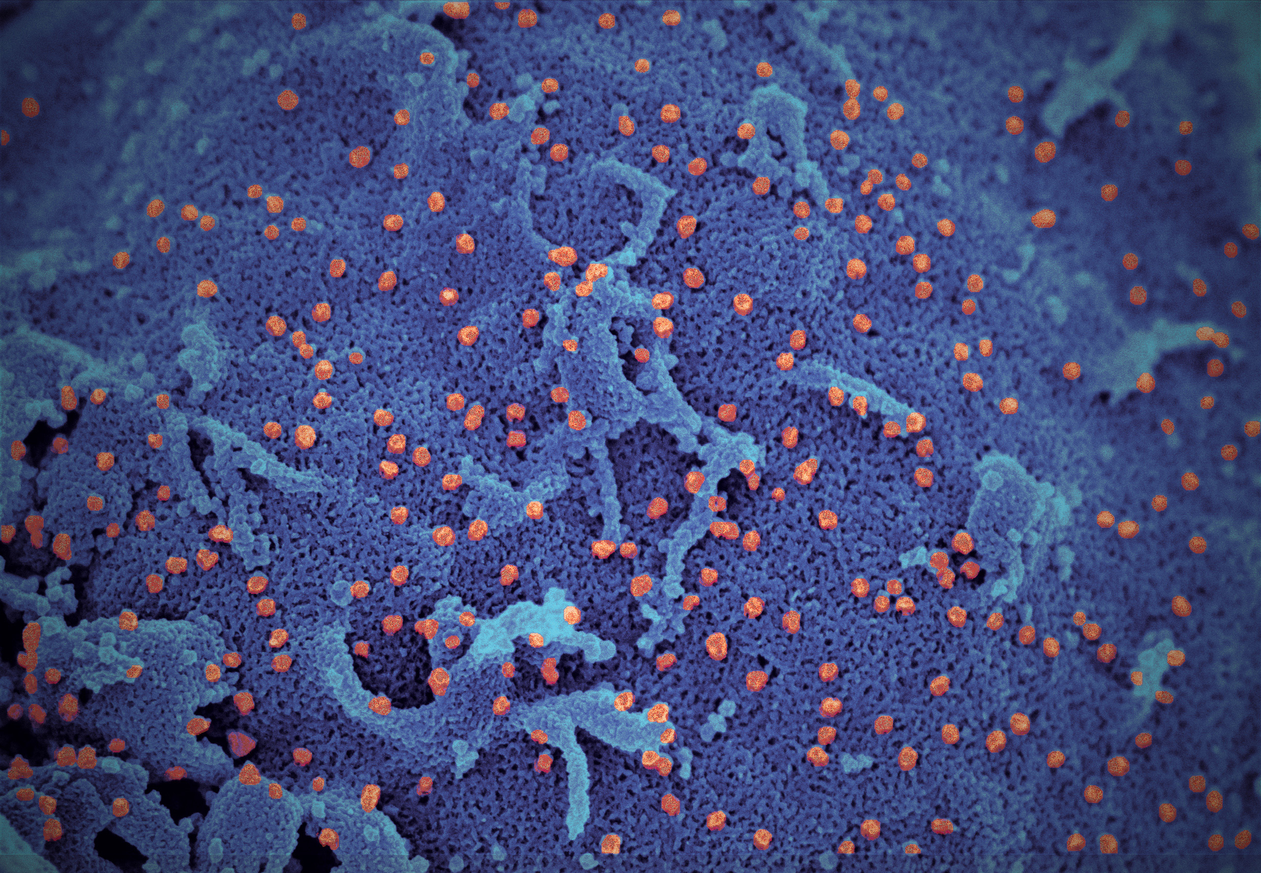
Pseudo-colour scanning electron micrograph of SARS-CoV-2 grown in culture from a patient isolate. After 24 hours in culture there are large numbers of viral particles (orange) on the surface of the cell (blue).
Image credit: LKS Faculty of Medicine, Department of Electrical and Electronic Engineering (K.Tsia, K.Lee and Q.Lai), and Electron Microscopy Unit, The University of Hong Kong.jpg?h=1152&iar=0&w=2304&hash=658331D0AEAEDC391E852C233CB28345)
(Left) Low magnification electron micrograph of a monkey kidney cell (Vero E6) after infection with the SARS-CoV-2 Omicron variant showing cell damage with swollen vesicles containing small black viral particles.
(Right) High magnification electron micrograph of an infected Vero E6 cell showing aggregates of viral particles with corona shaped spikes on their surface (red box).
Image credit: Professor John Nicholls, Clinical Professor of Department of Pathology; and Professor Malik Peiris, Tam Wah-Ching Professor in Medical Science and Chair Professor of Virology, School of Public Health, HKUMed; and Electron Microscope Unit, HKU.
(Left) Low magnification electron micrograph of a Vero (monkey kidney) cell 24 hours after infection with the Omicron variant of SARS-CoV-2. The boxes show clusters of viral particles in the cytoplasm.
(Centre) High magnification electron micrograph of the larger box in the left panel showing aggregates of viral particles in vesicles and on the surface of the cell.
(Right) High magnification electron micrograph of the smaller box in the left panel showing large aggregates of viral particles with spikes (box) in membrane bound vesicles.
Image credit: Professor John Nicholls, Clinical Professor of Department of Pathology, HKUMed; Professors Malik Peiris, Tam Wah-Ching Professor in Medical Science and Chair Professor of Virology, School of Public Health, HKUMed; Professor Leo Poon Lit-man, Professor and Head of Division of Public Health Laboratory Sciences, School of Public Health, HKUMed; and Electron Microscope Unit, HKU.