Keynote Lecture III
HCC immune atlas by CytoF and PD1 trials experience in the Mainland
Fang Weijia
Zhejiang University
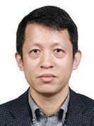
 Professor Weijia Fang is Director of Biotherapy Center of Medical Oncology, First Affiliated Hospital, School of Medicine, Zhejiang University. He has completed the rotating internship and residency at the First Affiliated Hospital, Zhejiang University and was subsequently awarded clinical fellowships in medical oncology in the same hospital. He established his own department and became Director of Biotherapy Center of Medical Oncology at the First Affiliated Hospital, Zhejiang University in 2014. He was elected a full member of American Society of Clinical Oncology (ASCO) and the Chinese Society of Clinical Oncology (CSCO). He has accumulated a wealth of clinical experience in the chemotherapy, target therapy and biotherapy of solid malignancies, mainly in digestive system neoplasms. He has participated in more than 20 international and domestic clinical trials of anticancer therapies. His team focuses on the translational research of microflora and immune altas of the so-called Immuno-Oncology area.
學歷:
醫學博士
職稱,職務:
副主任醫師,科主任,碩導
醫院及科室:
腫瘤細胞生物治療中心、浙江大學醫學院附屬第一醫院
學術兼職及交流:
美國印第安那大學 訪問學者
香港大學 訪問學者
香港大學鄭裕彤博士獎助金獲得者
中國臨床腫瘤學會青年專家委員會 委員
歐美同學會 會員
CRHA精准醫學與腫瘤MDT專業委員會 常委
CRHA胰腺疾病專業委員會 委員
中國醫師協會整合腫瘤學專委會 常委
中國醫藥生物技術協會精准醫療分會 委員
中國醫促會神經內分泌腫瘤分會 委員
中國抗癌協會腫瘤營養支援學組 委員
中國抗癌協會腫瘤轉移專業委員會 青年委員
浙江省醫學會營養與代謝分會 委員
浙江省醫學會腫瘤學分會 委員
研究領域及專業方向:
腫瘤基因大資料庫指導下的化療、靶向以及細胞生物免疫綜合治療
特長:
消化道腫瘤圍手術期腫瘤內科綜合診治
Abstract
Our team have accomplished the first part of our project of the mass spectrometry and the in situ single molecule experiments. The results are: (1) We preliminarily divided immunocytes in the liver into 33 subgroups by doing the mass spectrometry and the biological information analysis revealing a different and unmet atlas of T cells and DC cells subgroups, Monocyte/Macrophage cells, NK cells and B cells subgroups, et; (2) We had used the in situ single molecule dynamic technology to test the interaction of PD-1 and PDL-1 on the surface of T cells, and found by the first time to our knowledge that the biological mechanical force can extend the combination time of PD-1 and PDL-1, which potentially regulated the PD-1 suppressing function. We suppose that the liver tumor microenvironment could change the modification state after translation of PD-1 on T cells or Kuppfer cells, which affect the dynamic interaction of PD-1 and PDL-1, and then suppress the activation of immune cells, leading to the occurrence and development of tumor.
團隊前期已進行初步質譜流式和單分子原位動態的實驗操作,預實驗結果如下:(1)通過質譜流式技術及生信分析,初步將肝臟組織免疫細胞分為33個亞群,其中有多種T細胞亞群、多種DC細胞亞群、多種Macrophage/Monocyte細胞亞群、多種Granulocyte細胞亞群、多種NK細胞亞群、多種B細胞亞群等;(2)採用單分子原位動態檢測技術,對T細胞表面的PD-1與PDL-1的相互作用進行了初步檢測,發現生物機械力能夠增強PD-1與PDL-1的結合時間,潛在性對PD-1的抑制T細胞的功能起到調控作用,這個發現是以往各項研究都沒有涉及過的。我們猜想肝膽腫瘤微環境有可能改變了T細胞上PD-1或者Kuppfer細胞上的PDL-1的翻譯後修飾,從而影響了PD-1/PDL-1動態相互作用的特性,從而抑制了免疫細胞的活化,導致腫瘤的發生和發展。


